Periapical radiographs provide important information about the teeth and surrounding bone. Some further techniques to make this procedure more comfortable for the patient include.

Periapical Radiography Pocket Dentistry
6 Which exposure technique does the American Academy of Oral and Maxillofacial Radiology and the American Dental Education Association recommend.

. The film is placed parallel to the long axis. Periapical X-rays are used to detect any abnormalities of the root structure and surrounding bone structure. The central rays of the x-ray beam could either be directed perpendicular to the long axes of the teeth Or perpendicularly to the plane of the image receptor but not perpendicular to both at the same time.
A periapical view shows the tooth from the occlusal surface or incisal edge to the. The paralleling technique is recommended for routine periapical radiography but there are. Occlusal X-rays are larger and show full tooth development and placement.
The film is placed parallel to the long axis of the tooth in question and the central x-ray beam should be directed perpendicular to the long axis of the tooth. The patient was positioned upright with hisher mouth was opened as wide as possible to allow the X-ray beam to pass to the sensor unobstructed from the opposite side of the mouth. The paralleling technique results in good quality x-rays with a minimum of distortion and is the most reliable technique for taking periapical x-rays.
The Bisecting Angle Technique is an alternative to the paralleling technique for taking periapical films. Paralleling technique Bisecting angle technique Paralleling technique It is also called the extension cone paralleling technique right angle technique and long cone technique. What is labial mounting.
An intraoral full-mouth survey FMX on an adult consists of _____ images. Periapical lesions a c ommon dental disease are conventionally detected by dentists through radiog raphy. Gently softening the edges of the film by rounding it very carefully between the fingers.
Bone about 2 to 3 mm beyond the apex. The X-ray head is directed at right angles vertically and horizontally of both the tooth and the image receptor. This study aimed to assess the application of.
Periapical Lesion Diagnosis Support System Based on X-ray Images Using Machine Learning Technique. The paralleling technique results in good quality x-rays with a minimum of distortion and is the most reliable technique for taking periapical x-rays. Each X-ray reveals the entire arch of teeth in either the upper or lower jaw.
By using a film sensor holder with still. Periapical X-rays show the entire tooth from the exposed crown to the end of the root and the bones that support the tooth. The extraoral periapical radiographic technique was performed for both maxillary and mandibular teeth using Newman and Friedman technique2.
Ensure they are seated high enough so it is easy to see the occlusal. The X-ray tubehead is then aimed at right angles vertically and horizontally to both the tooth and the image. 4 Which technique is most often used when exposing a periapical image.
To take a periapical exposure the hygienist or x-ray technician places a small photosensitive imaging plate coated with phosphorus into a sterile wrapper and inserts it into the patients mouth just like a conventional X-ray film card. With this technique the film is placed parallel to the long axis of a tooth allowing the X-ray to be focused perpendicular to the long axis of the tooth. 3 What is an intraoral technique of exposing periapical images.
These X-rays are used to find dental problems below the gum line or in the jaw such as impacted teeth tooth fractures abscesses tumours and bone changes linked to some diseases. And routinely used for investigating the periapical and. The accelerated x-rays are decelerated by the target material resulting in bremsstrahlung.
Bisecting angle and paralleling. Periapical film is held parallel to the long axis of the tooth using film-holding instruments. DENTAL X-RAYS X-rays are produced by boiling off electrons from a filament the cathodeand accelerating the el to the target at the anode.
In the paralleling technique the central ray of the x-ray beam must be ___ to the film sensor and the long axis of the tooth. Vo TN Ngoc 1 Do H Viet 2 Le K Anh 3 Dinh Q Minh 4 Le L Nghia 5 Hoang K Loan 6 Tran M Tuan 7 Tran T Ngan 8 Nguyen T Tra 9. The sensor was placed on the.
PARALLELING LONG-CONE PERIAPICAL EXPOSURE TECHNIQUES GENERAL A long cone is used to take x-rays with paralleling exposure techniques. The image receptor is placed in a holder and positioned in the mouth parallel to the long axis of the tooth under. Parallel technique The image receptor is placed in a holder and placed in the mouth parallel to the longitudinal axis of the tooth under.
There are two types of techniques used for periapical radiographs. Most frequently used radiography is for the periapical which is performed by the bisecting Thus when considering the execution of the radiographic technique and the possibility of errors that occur during the exposure of X-ray image XR receptors it is important to identify those that occur more frequently. 1 Department of Pediatric Dentistry School of Odonto-stomatology Hanoi Medical University Dong Da Hanoi Vietnam 26 Department of.
Basically there are two techniques for taking periapical radiography. This must be done only slightly and the film must never be bent outright. Find the long axis of the tooth and then find the long axis of the image receptor as it is placed next to the tooth.
5 What is bisecting angle technique in dentistry. The patient is seated upright in the dental chair and should remove any removable dental appliances glasses or jewelry that could interfere with the X-ray beam. Periapical radiography is an important diagnostic aid.
Periapical radiography is a commonly used intraoral imaging technique in radiology and may be a component of your radiologic examination. What are periapical radiographs used for. By using a filmsensor holder with fixed image receptor and.
The X-ray is taken and the exposed plate is then loaded into a scanner or processor which reads the image. The bisecting technique may have to be used for patients unable to accommodate the film positioning device used in the paralleling technique. These patients may include adults with low palatal vaults and children.
The central ray is directed to pass at a perpendicular angle to both the tooth and the film. Bending the film produces an artifact on the image that may render the film unusable.

How Make Periapical X Ray

Periapical Radiography Pocket Dentistry
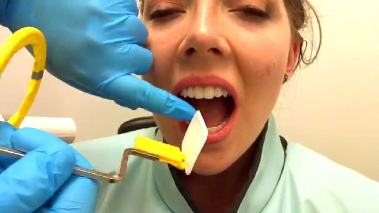
How To Take Periapical Radiographs Youtube
Periapical Radiography Pocket Dentistry

Periapical Radiography Pocket Dentistry

Periapical Radiography Pocket Dentistry

Periapical Radiography Pocket Dentistry

Periapical Radiography Pocket Dentistry
0 comments
Post a Comment